Introduction
Telemedicine, which has integrated two specialties, i.e. medicine and telecommunications, has enabled rapid electronic transmission of laboratory images (store-and-forward telemedicine) for various purposes: making initial diagnosis, obtaining a specialist’s second opinion, professional training and education, long-term data storage in digital format, and videoconferences (interactive application in real time) (1). Images are in laboratory medicine usually transferred as images taken by digital camera, and lately also as images obtained on microscopes with digital accessories. Digitalized images must have high resolution (2,3). In a complex process of digitalization, compression and electronic image transmission, the quality of the original image mustbe entirely retained. In order not to lose high resolution of an original image by compression, quality algorithms must be applied for image compression along with a high capacity telephone network (4). JPEG (Joint Photographic Expert Group) is a common name for a working group that has developed a procedure to compress still images. Development, evaluation and standardization were carried out from 1982 to 1987, and in 1992 the JPEG Group finalized the specification of procedures for lossy and lossless compression of tone still images that was accepted as a national standard (5). JPEG 2000 is a new international standard based on the wavelet technology (6). JPEG 2000 algorithm can entirely or partially code databases without data loss, which makes it suitable for processing of medical images (6). Regardless of what compression algorithm is used, it is important to determine the highest degree of image compression without loss in quality which is necessary for image evaluation (7). Image quality is rated according to subjective and objective criteria.
The aim of this paper was to compress the images of electrophoresis and isoelectric focusing samples by JPEG and JPEG 2000 algorithms, and assess the quality of compressed images before and after their electronic transmission. Electrophoresis and isoelectric focusing images had been used in a multicentric evaluation of patients with bisalbuminemia.
Materials and methods
Formation of digital images and compressions
Three images were selected for the study: a scanogram of cellogel electrophoresis of serum proteins of a patient with the Zagrebalbumin (Fig. 1.A1), two samples of polyacrylamide isoelectric focusing of the Krapina albumin (Fig. 1.B1), and a photograph of polyacrylamide isoelectric focusing of albumin samples with standards (Fig. 1.C1). The images were used in email correspondence related to publication of results of a study of double albuminemia (8,9). The resolution of original images was 1199 dpi (Figures A and B) and 99 dpi (Figure C). The selected images were scanned (using a flat scanner) and subsequently stored as computer images (the size of converted images was 225–649 KB; the number of pixels was 77–221 kilopixels). After storage, the images were converted to 24-bit BMP (BMP is a standard graphic format for MS Windows; folders with 24 bits per pixel are usually applied as this ensures better quality of the original image for polychromatic images) and compressed using VCDemo educational application, version 5.03 (10). During compression, two different algorithms for image compression were tested, i.e. JPEG (5) and JPEG2000 (6). Electropherograms were compressed at eight different degrees by both compression methods, i.e. at 3.0 bpp (bits per pixel), 1.5 bpp, 1.0 bpp, 0.7 bpp, 0.5 bpp, 03 bpp, 0.2 bpp and 0.1 bpp. Finally, original and compressed images were emailed as attachments to different persons.
Quality measures
The quality of compressed and emailed images was tested in a subjective and objective manner: (i) three original images (Figs 1.A1, 1.B1, 1.C1) and all compressed images (3 x 17 = 51 images) were examined. The images were rated by eight medical biochemists from five different institutions: six from Croatia, one from Italy (L.M.) and one from Denmark (U.K.-H.). Except for L.M. and U.K.-H., others were not aware of the algorithm used for image compression. All assessors were familiar with the elements important for assessing image type, and they were asked to compare the compressed images with the original ones and rate them as excellent (of equal quality to the original image; unaltered), good or moderately blurred (slightly changed), or completely useless (total image degradation; loss of relevant data; (ii) uncompressed and compressed images were objectively rated by independent assessors M.D. and S.G. who were aware of the protocols used for image compression.
The rating of images was carried out before and after compression by using software for image analysis that can determine signal-to-noise ratio (SNR), peak signal-to-noise ratio (PSNR), mean squared error (MSE) and optimized quality factor (OQF). Among the above parameters, MSE and PSNR are the most often applied measures of image quality (11). PSNR, SNR and MSE are realistic, objective image quality measures (11). The quality factor determines how much data is to be lost, which is directly reflected in the size of a compressed image (12). VCDemo application of the JPEG compression algorithm (10) enables determination of OQF, i.e. the smallest quality factor that yields an image with the highest similarity to the original image. SNR implicates a ratio between medium signal power and effective noise power, and is calculated by applying the following equation:
 (1)
(1)where A represents root mean square amplitude. Asignal and Anoise are effective values of signal amplitude and of noise amplitude.
MSE is calculated according to the following equation:
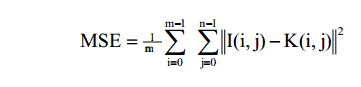 (2)
(2)where I (i,j) is the pixel amplitude of the original image, and K (i,j) is the pixel amplitude of the reconstructed image. M and n parameters represent the number of elements of an image in horizontal and vertical direction (expressed in pixels).
PSNR is defined as:
 (3)
(3)where MAXI is the maximum possible pixel value of the image, and MSE is the mean squared error (squared sum of rater bias and variance).
Statistical significance was tested by Wilcoxon-Mann-Whitney two-sample test, and the correlation between two variables was expressed by the coefficient of correlation (r). Cut-off values of OQF, PSNR and MSE were determined using ROC analysis (13).
Results
Subjective rating
Figures 1.A1, 1.B1, 1.C11 and their compressed variants were transmitted as email attachments to eight local and international experts. The recipients agreed that all images compressed by JPEG2000 algorithm were excellent, i.e. they were of the same quality as original images. On the other hand, JPEG compression was rated as less useful (Table 1). Only the images compressed at 0.3-3.0 bpp were rated as excellent. Compressions at 0.2 bpp were rated as good, and compressions at 0.1 bpp resulted incompletely useless images. Loss in quality of images compressed at 0.2 and 0.1 bpp was present already before electronic transmission (compare images 2 and 3 to images 1 in Fig. 1). The transmission itself did not affect image quality (not shown).
Table 1. Subjective evaluation of images at different JPEG compressions
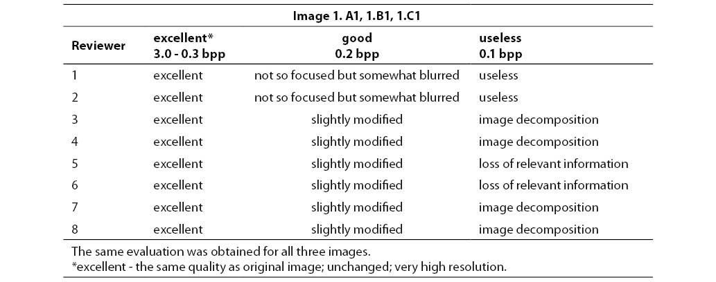
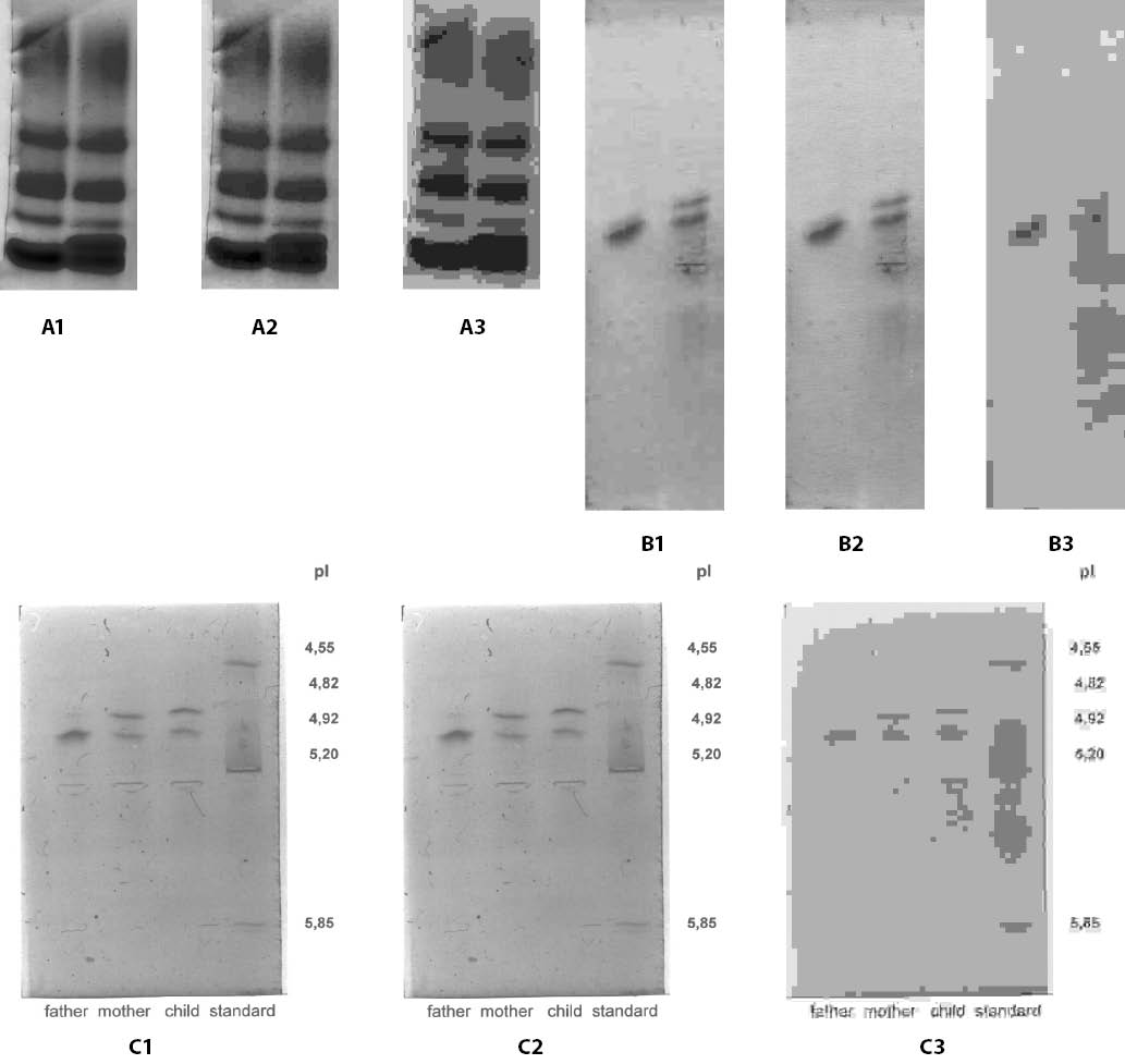
Figure 1. (a) Serum protein electrophoresis on cellogel (normal serum (left) and serum containing the slow-migrating albumin Zagreb (right); 133 kilopixels; 391 kB at JPEG-compression degrees of 3.0 bpp (A1), 0.2 bpp (A2) and 0.1 bpp (A3). (b) Isoelectric focusing on polyacrylamide (normal albumin without (left) and with (right) the fast-migrating albumin Krapina; 77 kilopixels; 225 kB at JPEG-compression degrees of 3.0 bpp (B1), 0.2 bpp (B2) and 0.1 bpp (B3). (c) Isoelectric focusing of albumin on polyacrylamide (father-normal, mother and child with both normal albumin and fast-migrating albumin Krapina; 221 kilopixels; 649 KB at JPEG-compression degrees of 3.0 bpp (C1), 0.2 bpp (C2) and 0.1 bpp (C3). The pI-value of 4.82 represents the albumin variant, whereas the remaining values represent protein standards.
Objective measures of quality
Table 2 shows objective measures of quality for 48 images compressed by JPEG and JPEG2000 algorithms. Again, image transmission did not have any impact on their quality. PSNR values did not significantly differ between images compressed at ≥ 0.3 bpp. In contrast, a statistically significant difference was observed in SNR values for each image (P = 0.029), i.e. the SNR value depended on the type of the analyzed image (image content). However, there was no statistically significant difference for SNR values between JPEG and JPEG2000 compression algorithms (P = 0.797). Finally, the MSE values calculated for corresponding bpp values significantly differed (P = 0.037) between the two compression formats.
Table 2. Quality measures for images 1.A1, 1.B1 i 1.C1 compressed by JPEG or JPEG2000 algorithms
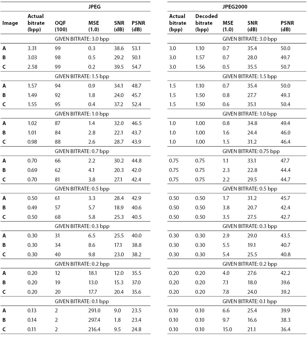
JPEG compression at 0.3 bpp and JPEG2000 compression at 0.1 bpp did not result in the loss of relevant data either for electrophoresis or isoelectric focusing samples regardless of the method of image formation. The images compressed by JPEG algorithm and rated as excellent had OQF values ranging from 31 to 99 (mean value 74; cut-off > 20). There was an overlap of PSNR values between images (JPEG compression) rated as excellent and images rated as good/useless. All images received as excellent had PSNR values > 37 dB (cut-off value) (Fig. 2). The values of PSNR (mean value 39.0 dB) and SNR (mean value 21.9 dB) at JPEG compression at 0.3 bpp could be compared to the values of PSNR (mean value 38.2 dB) and SNR (mean value 21.0 dB) obtained at JPEG2000 compression at 0.1 bpp. MSE cut-off values for images compressed by JPEG algorithm that were rated as excellent were ≤ 9.8. At the same bpp values, mean MSE values were 8.3 and 10.4 for JPEG and JPEG2000, respectively.
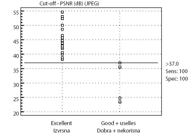
Figure 2. Cut-off value for PSNR at JPEG compression. All images accepted as excellent had a PSNR-value > 37dB (sensitivity 100%), while all images accepted as good or useless had a PSNR-value ≤ 37dB (specificity 100%).
Sens: 100 - Sensitivity 100% = 100% probability that PSNR will be > 37dB when images are evaluated as excellent.
Spec: 100 - Specificity 100% = 100% probability that PSNR will be ≤ 37dB when images are evaluated as good or useless.
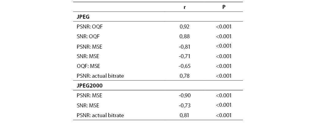
In JPEG compression, both PSNR and SNR correlated positively to OQF, and negatively to MSE (Table 3). PSNR was in positive correlation to the actual JPEG and JPEG2000 compressions. In JPEG2000 compression, PSNR and MSE, and SNR and MSE showed mutually negative correlation. All these negative correlations are theoretically based since MSE is an error measure, while PSNR and SNR are measures of quality (Equation 3).
Table 3. Correlation coefficients (r) for objective quality measures of images compressed by JPEG and JPEG2000 algorithms
Discussion
All images compressed by JPEG2000 algorithm were subjectively rated as excellent. In contrast, JPEG compression at 0.1 bpp resulted in images rated as completely useless; images compressed at 0.2 bpp were rated as good or moderately blurred, while those compressed at 0.3-3.0 bpp were rated as excellent. Higher degreesof compression with less distortion can be achieved using JPEG2000 algorithm than by, e.g., using JPEG algorithm. Clinically, subjective evaluation of image quality is of primary significance. Regarding objective evaluation of image quality, it is not easy to find objective numerical measures that would be useful for all compression methods (7).
PSNR, SNR and MSE are objective measures of image quality that are applied most frequently (11). In this study, a statistically significant difference was observed between MSE values determined for some images and MSE values calculated for the two compression schemes. According to Veldhuizen (15), MSE depends on the scaling of image intensity. PSNR values of different images did not differ significantly. As PSNR does not depend on signal power (unlike SNR), it shows the amount (power) of noise that affects an image. Also, since MSE is an error measure while PSNR and SNR are quality measures, their interrelationship was approximately inversely proportional so that MSE values were negatively correlated to PSNR and SNR. Therefore, SNR values were dependent on the type of the analyzed image. However, no statistically significant difference was observed between the SNR values obtained for the two compression schemes. It is interesting that our previous investigations of the quality of x-ray images demonstrated that only PSNR values for digitalized images were higher than mean values of digital photos, both for JPEG and JPEG2000 (16). In this study, we noted that JPEG compression at 0.3 bpp and JPEG2000 compression at 0.1 bpp did not result in the loss of relevant data either in electrophoresis or isoelectric focusing, regardless of how an image was obtained. It is important to point out that PSNR and SNR values (39.0 and 21.9 dB, respectively) at the JPEG compression rate of 0.3 bpp corresponded to PSNR and SNR values (38.2 and 21.0 dB, respectively) obtained for JPEG2000 compression at 1.0 bpp. The corresponding MSE value was 8.3 for JPEG and 10.4 for JPEG2000. All images rated as excellent had the PSNR value > 37 dB, and all images received as good or useless had PSNR value ≤ 37dB (Fig. 2). This PSNR cut-off value may be applied to distinguish high quality- from low quality images. Still, PSNR cut-off values are, according to Ashraf et al. (17) different for different images (retinal image: 30.2 dB; angiogram: 38.3 dB; lung x-ray: 31.2 dB, all for JPEG algorithm) (17). Yamamoto et al. (18) showed that PSNR values for high quality CT lung images ranged from 45.3 to 44.1 dB. In a previous study, PSNR cut-off values for digitalized lung x-rays were 45.3 dB for JPEG at 0.3 bpp and 44.1 dB for JPEG2000 at 0.1 bpp (17,19). Accordingly, it appears that PSNR values depend on image content. Actually, the capacity of an adequate compression method is related to image content: an image with less details is easier to compress than an image with many details. Chandra et al. demonstrated QF to be a good indicator of the quality of images compressed by JPEG algorithm (20), and showed that OQF value of 75 stands for images of excellent quality, which is consistent with our study where mean OQF value was 74. OQF correlated positively with PSNR and SNR, and negatively with MSE. The results presented in Table 2 reveal that equal bit rate values yielded different OQF values (at 1.0, 0.7, 0.5, 0.3 and 0.2 bit rates), which is consistent with results by Fidler et al. (21) who showed the OQF value to be important for reproducibility of image details. One of the limitations of our study was certainly a low number of original images, which might have influenced statistical analysis results. In a potential future investigation it would be necessary to analyze a higher number of original images with different content and also choose some other objective measures to establish image quality. Additional limitation is the fact that quality assessors were not blinded to the elements that were important for assessing image type, which might have resulted in a bias in their subjective rating.
Precedence should be given to JPEG2000 compression algorithm, which was actually recommended by the National Electrical Manufacturers Association that included JPEG2000 among DICOM (Digital Imaging and Communications in Medicine) standards for compression of medical images (22). If JPEG standard is applied, care should be taken of the highest degree of compression that can be used to maintain image quality at the same time. In Croatia, interest in telemedicine is focused on virtual e-medical centers (23), e-health care centers and island medicine (24), and telesurgery (25). Recommendations for online communication in telehematology and other telemedical consultations are available in the Rules on Implementing Telecommunication Consultations (only in Croatian) (26). As international uniformity of methods in telemedicine progresses, we suggest that, except in telemicroscopy, JPEG compression algorithm is, after assessment of a substantial number of images of different content, also used for image filesproduced in a medical biochemistry laboratory.
Acknowledgments
We hereby express our gratitude to medical biochemists of the Srebrnjak Children’s Hospital, of the Clinic for Tumors, and of the Department of Medical Biochemistry and Hematology, Zagreb University School of Pharmacy and Biochemistry, for their participation in this study.