Introduction
Urinalysis is the physical, chemical, and microscopic examination of the urine. It encompasses a number of tests to detect and measure cellular or biochemical elements that might be present in this biological fluid. As in other areas of laboratorydiagnostics, however, a compelling and critical requirement is to achieve improved standardization of performance, not only for an essential clinical need (i.e., adoption of consistent reference intervals and appropriate interpretation of results), but also because urinalysis continues to be one of the most frequently laboratory tests requested in clinical laboratories (1). Urinalysis is in fact strongly recommended in the presence of:
1. suspected urinary tract infection;
2. suspected (or follow up of) non-infectious disorder of the urinary tract primarily due to systemic diseases such as rheumatic diseases, hypertension, toxaemia of pregnancy;
3. suspected (or follow-up of) kidney disease;
4. suspected (or follow up of) non-infectious renal disease and recurrent urinary calculi;
5. diagnosis or monitoring of side effects of drugs (1).
It is instead discouraged in the settings of patients with diabetes mellitus, where metabolic control with glycosylated hemoglobin is preferred, and in pregnant women who can be more reliably monitored with proteinuria using specific quantitative assays (1).
Urinalysis has undergone radical changes over the past few years, so that the extra-analytical phases of this test should also be revised according to the most recent technical advances (2). The current recommendations for the appropriate performance of the test are retrievable from the European Confederation of Laboratory Medicine (ECLM) – European Urinalysis Guidelines, a consensus document published in 2000, which objective is “try to create a practice of consensus for urinalysis”. This guideline takes into account the whole process of urinalysis, including the preanalytical phase, the clinical aspects supporting the appropriate choice of the tests, the diagnostic algorithms, the production of a suitable laboratory report and the appropriate interpretation of test results. Although it still represent the mainstay in urinalysis, this guideline should however be reassessed according to the recent developments taking place in preanalytical materials and analytical techniques (e.g., new methods of collection, storage, handling, transportation and analysis of the specimens), as well as according to the renewed clinical reasons underlying the request of this essential diagnostic test.
The specimen
Several lines of evidence attest that the first-void urine morning sample might be preferred for chemical analysis inasmuch as this specimen usually contains an appropriate number of the elements to be analyzed. This specimen is also suitable for microbiological investigations (1). It is also recommended that the early morning urine be voided after an 8-h period of recumbence and after not less than 4 h storage time in the urinary bladder (even if the bladder was emptied earlier during the night). When collecting the sample, the first portion of the urine should be discarded, since it is frequently contaminated by the commensally urethral flora, in both genders. The mid-stream urine appears to be the most suitable also for physical, chemical and morphological analyses, since it is minimally contaminated by urethral secretions of mucus. In special circumstances, such as accurate analysis of cell type and morphology for diagnosing glomerular or not glomerular haematuria, atypical transitional or squamous cells, decoy cells due to polyomavirus and Bacillus subtilis (BK) infection, a second void specimen might be preferable, since the permanence of urine in the bladder might compromise the shape and even the integrity of some cellular elements (1,3).
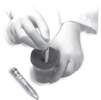
Figure 1. Urine specimen containers using a closed, vacuum device.
Sample collection
Before collection of a suitable urine sample, it is advisable to inform the patient on the reasons for performing the test. Comprehensive instructions should be provided on the appropriate procedure for collection. Ideally, these instructions should be given both orally and in written form, possibly accompanied by figures and illustrations, which would make the entire process more easily understandable to everybody. According to the European Urinalysis Guidelines, the entire genital region should be carefully washed with water. As mentioned, the first part of the urine should be eliminated and the midstream should be collected in a sterile container (1). The container should then be closed with a hermetical seal and sent to the laboratory as soon as possible (1,4). The recent availability of secondary and sterile vacuum tubes makes this process simpler, since they allow the transfer of an appropriate amount of urine for physical, chemical, morphological and microbiological examination without opening the original container (5). Devices with a wide base to avoid accidental spillage and that can be easily capped are preferable, since they allow transportation of the specimen with a minimum risk of leakage. The use of sterile containers with airtight cap and luer adapter predisposed to be connected with secondary vacuum tubes for direct, fast and safe transfer of urine specimen to evacuated tubes via vacuum pressure have several practical, technical and clinical advantages (Figure 1), including a more convenient and standardized approach of urine collection, a lesser likelihood of contamination, the possibility to use the same and therefore comparable and equally representative material for both standard and microbiological testing, along with a significantly lower biological risk for the healthcare personnel, who can directly transfer the material from one container to another (1,5,6).
Sample handling
The procedures to be followed immediately after sample collection include the appropriate labeling of the specimen (e.g., the presence of patient’s data, specimen type – first or second voiding of the morning, random sample – time and method of collection, and complementary information such as the presence of preservatives and the relative hazard symbols). The label should have an appropriate placing, allowing a clear view of the content. It should hence be placed on the container and not on the cover. In the case the sample is to be transferred (e.g., to a core laboratory from a peripheral center), biohazard labels and packaging must comply with the current European Standard EN829 (1,6).
Sample storage
Various strategies are currently being implemented or advocated to contain and reduce the overall costs of laboratory services. These include centralization, consolidation and integration of services, reengineering of laboratories on large networks, increasing the level of automation, optimizing test usage, decentralizing testing with point-of-care devices. As such, delayed sample analysis might be a rather frequent circumstance in modern clinical laboratories, especially when samples are shipped from peripheral collection centers to distant core laboratories. Although urine analysis within a short time frame after collection is still the gold standard (1), urine samples stability and storage are becoming leading issues to be considered in the preanalytical phase of urinalysis, likewise other areas of in vitro diagnostics.
Storage at +4 °C is currently recommended when the analysis of the samples can not be performed within one hour from collection (1). This is however a very critical and debated issue, in that some recommendations have provided different and often conflicting indications. It seems however reasonable to store the samples at room temperature and with no preservatives when the analysis can be performed within 1 hour from collection, whereas refrigeration might be indicated in all the other conditions. A variety of preservatives have been recently developed and commercialized, with the aim to achieve urine stabilization before chemical, physical and microbiological analysis. Nevertheless, the use of these substances is still a matter of debate, because there are some evidences that the various additives might interfere with some tests (1,4,6,8–11). No clear indications are available as yet on how these substances might affect some key aspects of urinalysis, especially on the automatic identification and enumeration of urine elements (e.g., bacteria, mycetes, leukocytes, erythrocytes), so that the increasing use of additives should prompt an accurate investigations on their influence on the analytical performance of the different diagnostic systems locally in use (9,10).
The synthesis of the best practice encompasses thereby that a sterile container should be given to the patient upon arrival to the healthcare facility (which might be essential for microbiological examination), from which a secondary vacuum tube based on a closed sampling system can be obtained for physical, chemical and morphological analysis. From a technical standpoint, the increasing automation characterizing several phases of urinalysis should also prompt the diagnostic industry to develop further technical skills and manufacture analytical platforms with sampler aspiration devices which would be able to process primary vacuum tubes. This will substantially increase the efficiency (i.e., throughput) and quality of testing, contextually improving the safety of the entire process (5).
The postanalytical phase
Although the vast majority of errors still occur in the preanalytical phase of urinalysis, major efforts should also be focused on increasing the clinical significance of the laboratory report. This might be essential considering the enormous advances that have occurred in the urinalysis over the past 10 years, mostly due to the widespread introduction of automation of the physical, chemical and especially of the morphological analysis. Major areas of improvements include standardization and harmonization of test results reporting, as well as inclusion of new parameters provided by the new analytical platforms.
As regards data reporting, a further harmonization of practices among different laboratories (e.g., according to the the European Urinalysis Guidelines), should be pursued. As such, results should be reported from ordinal scale examinations (e.g., negative, 1z, 2z or 3z) and the report should include traceability of date, time, technique/instrument and operator ID. The range of concentrations for each parameter should be always reported and preferred over a single arbitrary concentration (i.e., “0.2 ± 1.0 g/L” versus “1z – 0.3 g/L”). To increase standardization, manufacturers should be encouraged to adopt the same arbitrary categories for rapid examinations. When reporting results from particle analysis, the method used should be clearly described in the lab report (e.g., standardized sediment, standardized chamber counting, and automated analysis). Furthermore, morphological details should be reported at a basic or advanced level, measure units should be defined (the recommended unit for publication is particles/L, so that particle count should be finally expressed as averages per unit volume and not as ranges). Micro-organisms and clumps of cells, which are virtually uncountable by scrutiny, should be reported in ordinal scale from, e.g., “negative” to “3z”.
Conclusions
Great focus has been placed on the quality of clinical chemistry and immunoassay testing over the past decades, and therefore urinalysis has been somehow neglected. Nevertheless, the extraordinary advances occurred in this test require urgent actions to intervene both upstream and downstream the analysis, identifying new means for collection and storage of the samples, innovative containers that enable standardization and improve the efficiency while maintaining the quality throughout the total analytical process and especially in the chemical, physical, morphological and microbiological examination. The most reliable approach to improve total quality in urinalysis encompasses patient information (e.g., by using illustrations and figures) on the clinical significance of the examination and the appropriate procedure for sample collection, as well as the dissemination of available guidelines and best practice recommendations, which would enable:
1. control and standardization of those preanalytical processes most vulnerable to uncertainty and errors;
2. definition of precise levels of preanalytical related information and data on the samples;
3. identification of criteria for sample acceptance and rejection;
4. sharing approaches for comments and notes of non-compliance included in the laboratory report; and
5. appropriate interpretation of laboratory reports, to make test results more usable and clinically effective.
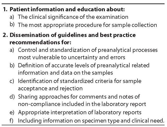
It is also to mention that something should be done to improve the pre-preanalytical phase, that is focusing on the appropriateness of the test request and attaining a variety of clinical and practical information (i.e., description of the specimen type and information of the clinical need) to assist the selection of examination procedures and the correct interpretation of test results (Table 1). Preferably, these data should accompany the specimen, e.g., being coded on the label on the sample tube.
Table 1. Major suggestions to improve quality in urinanalysis.