Leida Tandara [*] [1] Ilza Salamunic [1]

Show more about author

Introduction

 
Iron is essential element involved in a broad range of biologically important reactions critical for cellular function and also plays fundamental role in oxygen transferring. Disorders of iron homeostasis are among the most common human disorders. Although it is the fourth most abundant element in Earth´s crust, iron bioavailability is very low and despite a low daily requirements iron deficiency is the most common nutritional disorder in the world (2,3). On the opposite end of the iron disorders spectrum there are iron overload diseases which represent heterogeneous group of hereditary as well as acquired conditions. Hereditary hemochromatosis (HH) is a common inherited metabolic disease in people of Northern Europe (4). Excessive iron accumulation leads to the damage of liver, heart, pancreas and other organs. Beside systemic disorders of iron homeostasis local mismanagement of iron plays a role in several disorders. Neuronal disturbance of iron homeostasis and deposition of excessive iron in brain is associated with neurodegenerative disorders such as Parkinson´s disease, Alzheimer´s disease and Friedriech´s ataxia (5,6). Accumulation of mitochondrial iron is characteristic of sideroblastic anemia, erythropoietic protoporphyria and myelodisplastic syndrome (7). Furthermore, disturbed iron physiology mediated by proinflammatory cytokines and hepcidin leads to anemia of chronic disease (ACD) associated with various types of infections, hematologic malignancies, solid cancers, autoimmune disorders (8). Recently, a significant progress in understanding of iron metabolism has been made and new molecular participants have been characterized. Article gives an overview of the current understanding of iron metabolism: absorption, distribution, cellular uptake, release, and storage. We also discuss mechanisms underlying systemic and cellular iron regulation with emphasis on central regulatory hormone hepcidin.
 

Iron as double distress in living organisms

 
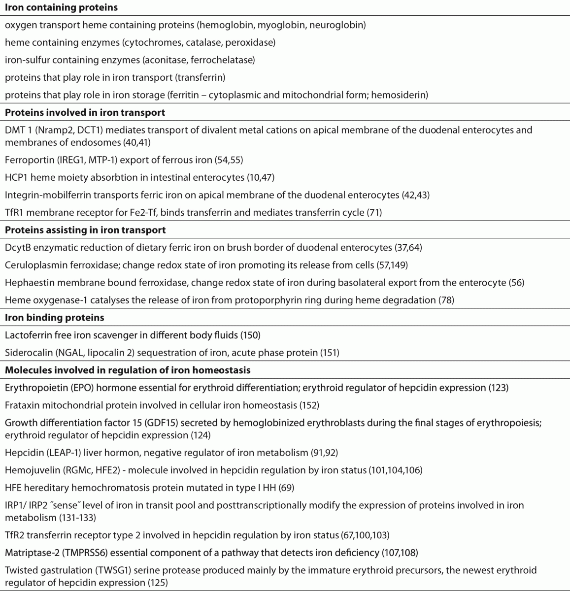
Iron is involved in a broad range of biologically important reactions. It is a component of innumerable hemoproteins including oxygen transport proteins, heme containing enzymes and many essential non-heme iron proteins that catalyse a wide range of reactions and have a central role in mechanism for oxygen sensing (Table 1) (9-11). Iron is a transition metal and it exists in two readily reversible redox states: reduced ferrous (Fe(II)) form and oxidized ferric (Fe(III)) form. At physiological oxygen concentrations the stable state of iron in most of its biological complexes is Fe(III) (12). Reduction reactions play a crucial role in the iron metabolism, because only reduced iron ion can be a substrate for transmembrane transport of iron, loading and releasing of iron from ferritin and for heme synthesis (12,13). Although biological function of iron is largely attributable to its chemical properties as a transition metal, exactly these properties render it potentially toxic. Iron excess is believed to generate oxidative stress due to its ability to generate ROS (7,14). Fe(II) catalysed reduction of one electron in O2 molecule results in the formation of superoxide anion which further leads to the sequence of well-known Haber-Weis-Fenton´s reactions that generate ROS which can possibly damage macromolecules such as proteins, lipids and nucleic acids (12).
 
Table 1. Key proteins involved in iron homeostasis.


Distribution of iron in the body

 
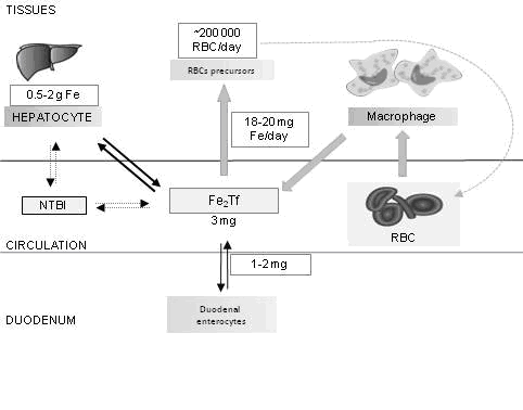
Body iron content of an adult is 3-5 g (~ 45 mg / kg woman, ~ 55 mg / kg for men). Most of the body iron is incorporated in hemoglobin of circulating erythrocytes (60-70%). Approximately 20-30% is in the form of ferritin and hemosiderin in hepatocytes and RES macrophages as a spare iron. While adult male has a 0.5-0.2 g of stored iron, children, adolescents and women of childbearing ages almost lack iron reserve. A small amount of residual iron in the body is in the form of myoglobin in the muscles or incorporated in enzymes (11). Amount of iron bound to transferrin is about 3 mg, but this iron transport compartment is very dynamic and changes about 10 times during the day (Figure 1).
 
Figure 1. Body iron distribution.
Most of the body iron is incorporated in hemoglobin of circulating erythrocytes (60-70%). Approximately 20-30% of iron in the body is in the form of ferritin and hemosiderin in hepatocytes and RES macrophages as a spare iron. The amount of iron bounded to transferrin is about 3 mg but plasma transferrin compartment functions as transit compartment through which flows about 20 mg of iron each day. Under circumstances of iron overload NTBI can appear in plasma. The bone marrow is the main consumer of circulating iron. 18-20 mg of iron, mostly recycled, is used for hemoglobin synthesis in 200 billion new erythrocytes every day. Healthy people absorb 1-2 mg of iron per day which compensates for iron loss.

NTBI - non-transferin bound iron; RBCs - red blood cells; Tf - transferrin.


Body iron stores

 
Body iron is stored in the liver, in both hepatocytes and Kupffer cells, in form of ferritin and hemosiderin. When cellular iron exceeds requirements the excessive iron is stored in bioavailable form as ferrritin which protects cells from potentially toxic reactions catalyzed by iron (15). Ferritin thus has got dual function of iron detoxification and reserve. It is composed of 24 subunits of two types: H (heavy or hart Mr ~ 21 kDa) and L (light or liver Mr ~19 kDa) forming a spherical structure. The ratio of H and L subunits within the assembled ferritin protein varies depending on tissue type. Incorporation of iron into ferritin requires ferroxidaze activity that is attributed to H subunit while L subunit has a role in mineralization (16). Mature ferritin has got a molecular weight of about 450 kDa and is capable to accumulate up to 4,500 iron atoms (12). Ferritin stored iron will be utilized when cell become iron deficient but mechanism underlying the ferritin iron release have yet to be completely elucidate (13,17,18).
Although the most of mature ferritin is located in the cytoplasm of cells, a small fraction was also found in nucleus of some cells. In the nucleus, ferritin could serve for delivering iron to iron-dependent enzymes or transcription factor activities but could also have a role of free iron "scavenger" that might otherwise catalyze DNA oxidative damage (19,20).
Also, human mitochondrial ferritin (MtF) is recently discovered. Mitochondria are organelles that exhibit a very high iron turnover carrying out the biosynthesis of heme and enzymes that contain Fe-S group. Since both mitochondrial iron deficiency and excess impair the metabolic and respiratory activities of the mitochondria, iron homeostasis within organelle must be strictly controlled. It is assumed that ferritin could play a role in storing iron within mitochondria protecting it from oxidative stress (21). MtF is expressed at extremely low levels in most cells. Studies have shown that MtF overexpression significantly affects intracellular iron homeostasis leading to a rapid redistribution of iron from cytosol to mitochondria where it is deposited in a form unavailable for metabolic use (22,23). Recent study indicates that up-regulation of MtF in erythroid progenitors may interfere with either the JAK2/STAT5 regulatory pathway or heme synthesis, contributing to ineffective erythropoiesis (24)
Although most ferritin is used to store iron within cells, very small amount enters into circulation. The source and detailed secretory pathway of ferritin are not completely understood but its biophysical characteristics imply active secretion through the lysosomal secretory pathway (25,26). Plasma ferritin is almost non-ferous, and its exact biologic purpose is still unknown. Some authors hypothesize that it may have a role as iron scavenger and modulator of inflammation (27). Studies have shown that extracellular ferritin can function as an iron carrier to provide iron to cells (28,29). Ferritin receptors are presented on lymphocytes and on some other cell types, but their physiological functions have not been fully defined (30). The plasma ferritin concentration is used as useful indicator of iron stores (31). It has been estimated that plasma ferritin concentration of 1 µg/L corresponds to 8-10 mg tissue iron stores (32).
Another form of stored iron in the cell is hemosiderin, insoluble degradation product of incomplete lysosomal degradation of ferritin. In iron overloading conditions hemosiderin becomes the predominant iron storage protein. Under physiological conditions hemosiderin is not an effective iron donor but plays a protective role. Subject to conditions such as inflammation and hypoxia it could become an iron donor promoting free radical production and tissue damage in iron overloaded cells (33).
 

Intestinal iron absorption

 
There is no known regulated pathway of iron excretion so body iron content is regulated by precisely controlled intestinal absorption. The intestinal mucosa responds to changes in body iron stores, tissue hypoxia, and demand for iron, and it alters absorption accordingly. Absorption is increased in iron deficiency while is reduced in the iron overload. Losses of iron from the body occur from desquamated epithelium of the skin, intestinal cells and intestinal secretions. An adult man loses only 1 mg of iron per day whereas women during reproductive ages lose twice that amount due to menstruation bleeding, pregnancy and childbirth. Healthy people absorb 1-2 mg of iron perday which compensates for iron loss. Iron requirements increase during adolescence due to growth and during pregnancy due to expansion of the blood volume and growth of the fetus (34). For optimal nutrition a daily intake of 8-10 mg of iron is required.
Dietary iron is present in food as one of two forms - as inorganic or heme iron. Inorganic form is dominant in the standard diet, and makes up about 90% of the total amount present in food. Heme accounts for only 10% of the dietary iron (35). Despite the relatively low participation in diet, heme is a highly bioavailable source of iron whose absorption is significantly more efficient than the absorption of inorganic form because it is not affected by the dietary constituents that adversely affect the absorption of inorganic form (36).
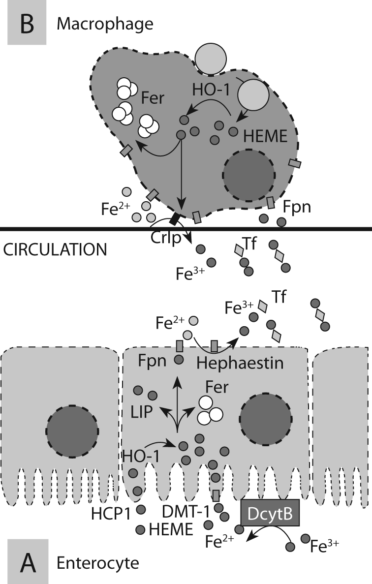
Absorption of iron is a very complex process that takes place in the duodenum and upper jejunum. This process involves number of proteins that transport iron across the apical membrane (importers), proteins that transport iron through the basolateral membrane of enterocyte (exporters), proteins that change redox state of iron and thus assist in its transport (Table 1; Figure 2A; Figure2B)).
 
 
Figure 2A. Absorption of iron in the gut.
Dietary iron could be absorbed as ferric, ferrous and heme iron. Ferric form must be first reduced to ferrous iron by DcytB. Fe(II) is then transported across the apical membrane into the cytoplasm of the duodenal enterocytes by DMT-1. Cellular uptake of ferric iron proceeds through a separate pathway, using IMT pathway while heme moiety is absorbed by intestinal enterocytes via HCP1. Within the cell iron is released from protoporphyrin ring by HO-1. After apical transport two forms of iron enter the inorganic iron pool of the enterocytes and could be sequestrated as ferritin or transported across the basolateral membrane. Iron basolateral export is carried out by ferroportin and also requires change of its redox state by ferroxydase – hephaestin. Ferric iron is captured by transferrin and distributed throughout the body.
 
Figure 2B. Recycling of iron by RES.
RES macrophages in the spleen and elsewhere phagocytize and lyse aged or damaged red blood cells. Heme is degraded by HO-1 and iron is liberated from protoporphyrin ring. Iron export by ferroportin requires also change of redox state and is accomplished by ceruloplasmin.

Crlp - ceruloplasmin; DMT-1 - divalent metal transporter 1; DcytB - duodenal cytocrome b; Fer - ferritin; Fpn - feroporrtin; HO-1 - heme oxygenase 1; HCP1 - heme-carrier protein; LIP - labile iron pool; NTBI - non-transferin bound iron; RBCs - red blood cells; Tf - transferrin.


Absorption of inorganic iron

 
The most dietary inorganic iron is in ferric form and it must be first reduced by brush border ferrireductase, duodenal cytocrome B (DcytB) (37). Some physiological approaches elucidate intracellular duodenal ascorbate as electron donor for this reductase. Recently, there are some data showing that members of six-transmembrane epithelial antigen of the prostate (STEAP) family are also expressed in intestine (38). The exact role of both types reductases especially in human iron absorption remains uncertain (39). Ferrous iron is then transported across the membrane into the cytoplasm via divalent metal transporter 1 (DMT1) expressed on apical membrane of the duodenal enterocytes (40). DMT1 is not specific for iron transport but also mediates transport of other divalent metal cations including zinc, manganese and copper although it seems that it’s primary physiological role is iron transport (12,40). This transport protein is also expressed on the membrane of endosomes where mediates iron transport from endosomes into the cytoplasm during transferrin cycle (9). DMT-1 seems to have a role in transport of non transferrin bounded iron (NTBI), especially in conditions of iron excess (41).
Some researches have shown that duodenal ferric iron uptake proceeds through a separate, although less understood pathway. While ferrous iron uses DMT-1, ferric uses integrin-mobilferrin pathway (IMT) that solely transports ferric iron, not other metals of nutritional importance (36,42). This pathway involves several proteins like mobilferrin, beta-3-integrin and flavin-monooxygenase. Flavin-monooxygenase has a role of ferrireductase. In the cell cytosol these proteins are integrated into a large protein complex called paraferritin (43). Western Blott analysis of paraferritin revealed that it also contains beta-2-microglobulin and DMT-1. The presence of considerable fraction of DMT-1, mobilferrin and hephaestin in the cytoplasm of cells indicates a possible intracellular role of these proteins (Figure 2A) (44,45).
 

Absorption of heme iron

 
Transport of heme across the membrane is not required only for heme iron absorption in gut but also for cellular heme turnover. Synthesis of heme partially takes place in the mitochondria and after that heme must be transported to the endoplasmic reticulum to be included in hemoproteines. Heme is involved in transcriptional regulation of some genes therefore also needs to be transported in the cell nucleus (46). Studies have shown that intact heme moiety is absorbed by intestinal enterocytes via heme-carrier protein 1 (HCP1), transport protein expressed at high levels in duodenum (47,48). Some recent investigation indicated that descripted heme intestinal transporter might be folate transporter (49). Within the cell iron is released from protoporphyrin ring by heme oxygenase 1 (HO-1) and converges with the cytosolic iron pool of enterocyte. Afterwards, two forms of iron share the same pathway - enter the inorganic iron pool of the enterocytes (Figure 2A). It seems that step catalysed by HO-1 is limiting factor in the absorption of heme.
 

Intracellular iron transport

 
Once iron enters the intestinal epithelial cells through the apical membrane, it could be sequestrated as ferritin or transported into circulation across the basolateral membrane. Absorptive enterocytes perform their function for two days and then are being shed into intestinal lumen. Iron that is not exported from enterocytes into the plasma is lost by exfoliation of intestinal epithelium. Therefore, transport of iron by ferroportin across the basolateral membrane determines whether iron is delivered into the circulation or removed from the body with shed enterocytes. Exfoliation of epithelial cells of intestinal mucosa may represent pathway of regulated iron excretion because these cells at the basolateral membrane express transferrin receptors type 1 (TfR1) and iron from the plasma can enter the cell by receptor mediated endocytosis, but the capacity of this mechanism of excretion in response to the accumulation of iron is very limited (50). To be transported through the basolateral membrane iron must first be transported through the cell cytoplasm. Transport of iron across the enterocyte cytoplasm is the least understood step in iron absorption. There are two possible mechanisms of transport that do not exclude each other: transport of iron associated with some proteins (chaperones) or transcytosis (51). Although the molecular details have yet to be explored, cytosolic monothiol glutaredoxins and Poly(rC)-binding proteins could act as iron chaperones and thus may represent the basic cellular machanism for intracellular iron delivery (52,53).
 

Basolateral iron transport

 
Ferroportin represents the only known iron exporter (54). This export protein is present in all tissues that export iron into plasma: basolateral membranes of duodenal enterocytes, membranes of RES macrophages, hepatocytes and placental cells. Mutations in ferroportin gene affect iron export from macrophages and cause type IV hemochromatosys (55). Iron basolateral export from the enterocyte requires also change of redox state by ferroxydase - hephaestin in the duodenum and ceruloplasmin elsewhere in the body, that converts intracellular Fe(II) back to extracellular Fe(III) (Figure 2A) (56,57). Ferroportin expression is increased in tissue macrophages as well as in enterocytes in response to iron-restricted erythropoiesis (58). Recent study provided evidence that erythroblasts express an important amount of ferroportin at their membrane. Authors propose hypothesis that in conditions of severe iron deficiency, the high ferroportin expression associated with low hepcidin levels could favour iron export from erythroblasts in order to abandon this essential element to cells more sensitive to iron deprivation (59).
 

Iron transport in plasma

 
The plasma transferrin compartment functions as transit compartment through which flows about 20 mg of iron each day (60). Principles of iron transport are partially dictated by its chemical properties. Binding of iron to transferrin, a major iron transporter in the blood, provide solubility, reduce reactivity and thus provides a safe and controlled delivery of iron to all cells in the body. Transferrin is serum glycoprotein with molecular weight about 75–80 kDa. Molecule is folded into two globular domains, each containing a specific binding site for single Fe(III). Affinity of transferrin molecule for iron at physiological pH is extremely high so almost all non-heme iron in circulation is bounded to transferrin (61). Under circumstances of iron overload NTBI appears in plasma. NTBI refers to all forms of iron in the plasma that binds to ligands other than transferrin. Iron is bounded to these ligands with substantially less affinity than to transferrin. This form of iron is very reactive and could enter Fenton reaction. NTBI is likely to be a major contributor to iron loading in hepatocytes under conditions of elevated transferrin saturation, capable to freely enter the cell so it is considered to be a marker of iron toxicity (62,63).
 

Iron uptake in the tissues

 
Transferrin is taken up into the cell by transferrin mediated endocytosis in so called transferrin cycle. Under physiological conditions this cycle enables controlled access of iron to cells because individual cells can efficiently regulate the entry of iron by regulating the expression of TfR1 at the surface, according to their iron needs.
 

Transferrin receptors

 
Two types of functionally different transferrin receptors are described, TfR1 and transferrin receptor 2 (TfR2). TfR1 is expressed by all iron-requiring cells but level of expression varies greatly (64). It is highly expressed on immature erythroid cells, rapidly dividing cells (normal and malignant), placental tissue. TfR1 is a transmembrane glycoprotein comprised of two identical disulfide bounded subunits with a molecular mass of approximately 90 kDa. Each subunit possesses one binding site for the transferrin. Diferric transferrin has a higher affinity for TfR1 than monoferric form or iron-free apotransferrin. Besides the membrane-associated TfR1, a soluble form of this receptor exists in human serum which represents a soluble fragment of the extracellular receptor domain. Soluble transferrin receptor (sTfR) is released by proteolytic cleavage of the protein C-terminal end. It is proposed that release of sTfR is regulated by binding of its ligand transferrin (65). The level of sTfR reflects the availability of functional iron.
TfR2 is predominantly expressed in liver, hematopoetic cells, duodenal crypt cells, and it overlaps with hereditary hemochromatosis protein (HFE). TfR2 binds to HFE and transferrin, but interacting domains of HFE with TfR2 are different from those of TfR1. It is assumed that TfR2/HFE complex is required for transcriptional regulation of hepcidin production by diferric transferrin (66-70).
 

Transferrin cycle

 
Binding of diferric transferrin to TfR1 at the surface of the cell triggers clathrin-mediated endosome formation and initiate transferrin cycle. Action of proton pump on endosome membrane acidifies endosome content and leads to a conformational changes of transferrin as well as transferrin receptor, resulting in iron release (71). Fe(III) is then reduced by ferrireductase STEAP3 and iron is transported across the membrane of endosome into the cytoplasm via DMT1 (72). Apotransferrin is then returned to the cell surface completing the transferrin cycle and is released to be recharged with iron. TfR1 is presented for a new uptake cycle. During its lifetime transferrin makes around 100-200 cycles of iron transport.
After cellular iron uptake iron enters poor characterized “labile iron pool“ (LIP). LIP is defined as pool of iron complexed with low affinity ligands (citrate, ATP, amino acids, ascorbic acid or by unidentified chaperones). Recent study identified iron(II)glutathione as the dominant component of this pool (73). LIP represents < 5% of the total cellular iron (74). It is dynamic compartment that supplies iron to the mitochondrion for heme and iron sulfur cluster synthesis or could be used for synthesis of iron-containing proteins in cytosole thereby controlling numerous metabolic reactions. Iron in the LIP that exceeds requirement for the synthesis of heme and non-heme iron containing proteins is stored within ferritin to minimize free iron because LIP is catalytically active and capable of initiating free radical reactions (75). Quantification of LIP is possible with novel nondisruptivetechnique that use fluorescent metalosensors and may be clinically significant in states of iron overload (76).
Under normal circumstances entry of transferrin bounded iron is the main route of iron entry into cells but the pathological accumulation of iron leads to transferrin saturation and appearing of NTBI which can enter into cells via transferrin-independent pathway (63)
 

Regulation of systemic iron homeostasis

 
Although iron is very abundant element in environment its bioavailability is very low so body uses iron very rationally. In the same time iron is potentially toxic and there is no pathway of regulated excretion so absorption in gut must be strictly controlled. Those facts dictate the principles of systemic iron homeostasis. The bone marrow is the main consumer of circulating iron and the most of the daily iron need is used for hemoglobin synthesis in 200 billion new erythrocytes. In balance, macrophages recycle 10–20 times more iron than the intestine absorbs providing most of daily iron supply. RES macrophages in the spleen and elsewhere phagocytize and lyse aged or damaged red blood cells. Heme is degraded by HO-1 and iron is liberated from protoporphyrin ring and released via ferroportin back to plasma transferrin (Figure 2B) (77,78). Changes in iron flux through macrophages affect the maintenance of iron homeostasis more rapidly than changes in iron absorption in enterocytes (79). Body losses 1-2 mg of iron per day and about the same amount is absorbed in gut in order to provide enough but not too much iron to keep stores replete. Therefore, systemic iron homeostasis regulates intestinal iron absorption, its entry and mobilization from the stores in order to meet erythropoietic needs. It also assures a stable milieu where each cell regulates iron uptake according to its own requirements.
Since its discovery many studies confirmed role of liver hormone hepcidin as key regulator of iron homeostasis and placed liver as the central organ of system iron homeostasis. This organ synthesizes hepcidin, main iron transport protein transferrin and stores the most of iron (80,81).
There is some evidence of kidney involvement in iron homeostasis at least when iron demand is high. Recently, several iron transport pathways have been identified in the kidney but the role of kidneys in iron metabolism should be explained by future studies (82-84).
 

Hepcidin

 
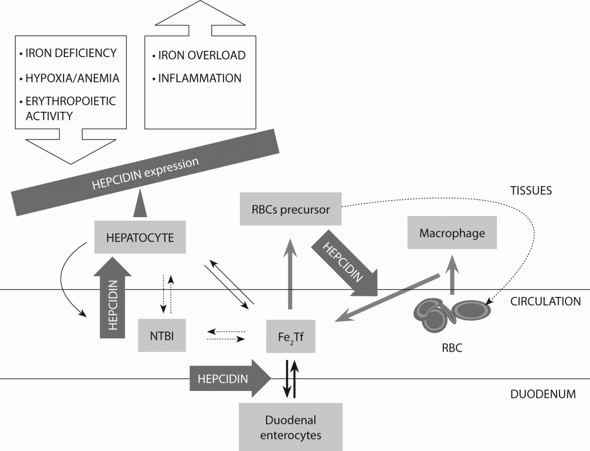
Hepcidin is negative regulator of iron metabolism. On the molecular level it binds to its functional receptor ferroportin promoting internalization, and finally lysosomal degradation of this iron exporter (85). Loss of ferroportin from cell membrane causes cellular iron retention and represses iron efflux from sites of main iron flow (macrophages, hepatocytes and enterocytes) into the blood decreasing thus transferrin saturation and reducing iron availability (Figure 3) (86).
 
 
Figure 3. Maintenance of systemic iron homeostasis by action of hepatic hormone hepcidin.

Liver cells receive multiple signals related to iron balance and respond by transcriptional regulation of iron regulatory hormone hepcidin. Hepcidin is negative regulator of iron metabolism that represses iron efflux from sites of main iron flow: macrophages, hepatocytes and enterocytes decreasing thus transferrin saturation and reducing iron availability. Iron deficiency, hypoxia/anemia and increased erythropoietic activity decrease hepcidin expression while iron overload (except HH) and inflammation increase it.

 
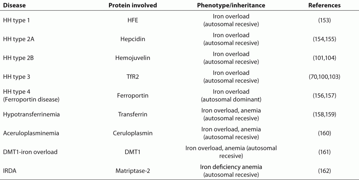
Table 2. Selected iron disorders of genetic origin.
 
 
Dysregulation of hepcidin production, whether genetic or acquired, causes iron disorder (Table 2). In healthy individual, an increase of body iron would lead to increased hepcidin expression and therefore to decreased iron absorption. In patients affected by HH, because of inadequate or ineffective hepcidin-mediated down-regulation of ferroportin, iron absorption continues despite high body iron load (87,88). Oppositely, overexpression of hepcidin gene is associated with a hypoferremic, microcytic, iron refractory anemia (89,90).
Hepcidin was isolated from human blood in the year 2000, during the searching for cysteine rich antimicrobial peptides. It is named LEAP-1 (liver expressed antimicrobial peptide) (91). Almost at the same time, a peptide was isolated from urine and named hepcidin after its hepatic origin and antimicrobial effect in vitro (92). Studies demonstrated that hepcidin is not liver specific but is also expressed in other tissues: the kidney, heart, lungs (93). Hepcidin is synthetized as an 84-amino acid (aa) prepropeptide, and is subsequently processed into 60–64-aa prohepcidin. Mature and biologically active 25-aa hepcidin is produced by the removal of the proregion with prohormon convertase furin (94). Hepcidin forms simple hairpin structure stabilized by four disulfide bonds (95). It also circulates in plasma as 22-aa peptide and as prohormone pro-hepcidin that lacks biological activity (93,96). Recent study revealed that proregion of hepcidin may have bacteriostatic effects, and as such may contribute to the innate immune response (97).
Hepcidin is transcriptionally regulated and there is no evidence of other control types. Numerous molecules are involved in regulation of its expression (Table 1). There are at least four major, separate pathways in hepcidin regulation: regulation by iron status, dietary iron and iron stores; regulation by inflammation; regulation by hypoxia/anemia; and regulation by erythroid factors (98,99).
 

Hepcidin regulation by iron status

 
Molecular mechanism by which iron stores regulate hepcidin synthesis is not completely described. HH caused by homozygous disruption of HFE, TfR2 and hemojuvelin (HJV) is characterized by low level of hepcidin inspite of iron overload indicating that these molecules act as direct or indirect regulators of hepcidin synthesis (100,101). At the subcellular level HJV and TfR2 are localized on the same basolateral membrane domain indicating that interaction of these proteins is possible. Localization enables direct contact with blood and sensing of signals influenced by iron metabolism (102,103).
HJV gene is identified in the year 2004 and its mutation is identified as a cause of type 2 hereditary juvenile hemochromatosis (HJH) (104). Patients with HJH mutations have very low urinary level of hepcidin and unlike other form of HH, early age of symptoms onset. HJV-mutant mouse exhibit severe iron overload phenotype and complete lack of hepcidin expression (101). HJV is expressed predominantly in skeletal and cardiac muscle and liver. This protein can be expressed as a membrane bound and soluble form (sHJV) detected in human plasma. It has been proposed that HJV act as co-receptor that binds to bone morphogenetic protein (BMP) ligands and BMP type I and type II receptors on the cell surface. This complex (HJV-BMP ligand-BMP receptors) consequently induces an intracellular BMP signalling pathway which in turn activates the SMAD4 signalling pathway. SMAD complex translocate to nucleus and directly increases hepcidin gene transcription (28,105). BMP/SMAD signaling cascade of HJV is important for basal regulation of hepcidin transcription (106). Recently, liver transmembrane serine protease, matriptase-2 (type II transmembrane serine proteinase; TMPRSS6) emerged as an essential component of a pathway that detects iron deficiency. It cleaves membrane bound HJV increasing sHJV that competitively impairs BMP signaling and blocks transcription of hepcidin gene permitting enhanced dietary iron absorption (107,108). Recent study presented data that do not confirm cleavage of membrane HJV by matriptase-2 in vivo suggesting that its role in hepcidin gene regulation could be more complex (109).
 

Hepcidin regulation by inflammation

 
Hepcidin is considered to be the mediator of ACD. Hepcidin synthesis is markedly induced by infection and inflammation. Interleukin 6 (IL-6) is apparently the key inducer of hepcidin synthesis during inflammation (110-112). It regulates hepcidin expression through signal transducer and activators of transcription (STAT3) signaling pathway (113). IL-6 acts on hepatocytes and stimulate hepcidin production resulting in cellular iron retention and hypoferremia, thus limiting iron availability to pathogens (112,114). Restricted iron availability is limiting factor in hemoglobin synthesis and results in development of anemia.
Almost all known bacterial pathogens require iron to multiply. Iron-withholding strategy is important component of innate immunity. Numerous studies confirm increased susceptibility to infection in patients with hemochromatosis due to increased iron availability (115). Decreased serum iron is believed to contribute to host defense against invading pathogens and cancer cells (116). In this sense hepcidin emerged as link between immunity and iron metabolism and the key mediator of anemia of chronic disease (117,118).
 

Hepcidin regulation by hypoxia/anemia and erythroid factors

 
When anemia/hypoxia occurs, erythropoietyn expression increases leading to stimulation of the erythropoiesis. In parallel, hepcidin gene expression decreases, allowing rapid mobilization of iron from reticuloendotelial cells and more iron is absorbed from the duodenal enterocytes to supply sufficient iron to erythrocyte precursor cells (119). Several studies indicated that suppression of hepcidin is not directly mediated by anemia or but requires tissue hypoxia increased erythropoiesis (120,121). Signals which regulate hepcidin expression are hierarchically arranged. In diseases characterized by ineffective erythropoesis, like talassemias, dominance of the stimulus of erythropoietic demand over the inhibition by iron stores can cause iron overload (122). Studies have shown that regulation of hepcidin by erythropoiesis is probably mediated by bone marrow-derivated signal molecules: growth differentiation factor 15 (GDF15), twisted gastrulation protein homologe 1 (TWSG1) and hormone erytropoetin (123-125). Suppression of hepcidin in hypoxia is mediated by hypoxia inducible factors (HIF) (126,127).
 

Regulation of cell iron homeostasis

 
Since both cellular iron deficiency and iron overload are detrimental for cell, iron uptake, storage, export and cellular distribution must be tightly controlled. Tight regulation of iron assimilation prevents an excess of free intracellular element that could lead to oxidative stress and damage of cellular structures like DNA, proteins and lipid membranes by ROS. At the same time it provides enough iron in order to meet the metabolic needs.
Cellular iron uptake and storage are coordinatively regulated at the posttranscriptional level by well-known IRE/IRP system. Cytoplasmic proteins known as IRP1 and IRP2 have the ability to ˝sense˝ level of iron in transit pool. This proteins bind specifically to RNA stem-loops known as IRE and posttranscriptionally modify the expression of proteins involved in iron metabolism.
IREs are stem-loop RNA motifs present on 3´ or 5´-untranslated mRNA regions (3´-UTR ur 5´-UTR) that can interact with IRP. Thus iron itself modulates the synthesis of variety of proteins involved in iron metabolism, heme synthesis, tricarboxylic acid cycle:
·        IRE at 5´-UTR mRNA   ferritin, ferroportin, cALAS, HIF-2-alpha;
·        IRE at 3´-UTR mRNA   TfR1, DMT1.
Recently, 35 novel mRNAs that bind IRP1 and IRP2 were identified as well as cellular mRNAs with exclusive specificity for IRP1 or IRP2 (128). Spontaneous mutations in IRE have been described in humans. Some genetic defects in L-ferritin IRE result in hyperferritinemia-cataract syndrome with prominent ocular findings and elevated serum ferritin but with no evidence of disturbed iron homeostasis. Mutations in H-ferritin IRE have been observed in cases with familiar iron overload disorder (129,130).
IRP1 is ubiquitously expressed cytosol iron-sulfur protein. When cellular iron concentration is sufficient IRP1 acts as an aconitase, cytosol iron-sulfur protein, and it lacks RNA binding activity (131). IRP2 functions only as an RNA binding protein which is degraded in the presence of iron but in the absence of iron it binds to IRE. Alternatively, genetic ablation of IRP1 and IRP2 revealed that IRP2 dominates iron homeostasis (132).
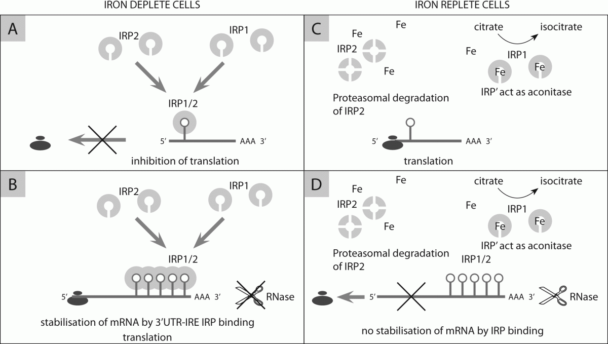
IRE-IRP binding has two different effects depending on the IRE location relative to coding region. Binding of IRP to IREs in 3´-UTR region stabilize the transcript and prevents mRNA degradation thus increasing mRNA translation and protein synthesis (Figure 4B). IRP binding to IREs at 5´-UTRs transcript results in translational repression by precluding ribosome binding and interrupting protein synthesis (Figure 4A).
 
 
Figure 4. Maintenance of cellular iron homeostasis by IRE/IRP system.

In iron deplete cells binding of IRP1/2 at 5´UTR IRE of ferritin and ferroportin mRNA block translation hampering its initiation (A); Binding of IRP1/2 at 3´UTR IRE of TfR mRNA stabilize its transcript (B). In iron replete cells IRP1 act as aconitase and IRP2 is degradated by proteases so translation of 5´UTR IRE mRNA of ferritin and ferroportin is carried out undisturbed (C). In presence of sufficient iron there is no binding of IRP1/2 and stabilization of 3´UTR IRE transcript of TfR (D).

 
When cells are iron-sufficient, IRP1/2 lose their affinity for RNA binding, consequently ferritin synthesis is activated while TfR1 mRNA is degraded. Opposite, when intracellular iron concentration is low IRPs bind to IREs of ferritin mRNA at its 5´-UTR and block translation, whereas stabilize TfR mRNA by binding at 3´-UTR and thus increasing iron uptake by cell. Iron is neither the only modulator of IRP-1 activity nor level of IRP2. Besides iron, this regulatory system is influenced by nitric oxide, phosphorilation by protein kinase C, oxidative stress and hypoxia/reoxygenation and this provides a molecular basis by which agents other than iron can selectively modulate iron metabolism in cells and tissues (133).
 

Future directions

 
Although iron metabolism once seemed quite simple, discoveries of numerous new molecules involved pointed out its complexity. New insights on iron metabolism and its regulation at both system and cellular level have opened up new diagnostic and especially therapeutic options not only related to disorders of iron metabolism, but to considerably broader spectrum of conditions.
Potential medical applications include development of molecules that directly or indirectly modulate hepcidin expression for the treatment of acquired and hereditary iron overload, or the treatment of ACD (134,135). Chronic kidney disease (CKD) is also associated with increased hepcidin levels so hepcidin antagonist could be useful as a supplement to erythropoietyn therapy in anemic CKD patients, especially in erythropoetin resistance.
Expression of ferroportin and hepcidin seems to be important predictors of breast cancer clinical outcome. Low ferroportin expression in breast cancer tissue provide sufficient amount of metabolically available iron to malignant cells and is indicator of poor breast cancer prognosis (136). Cancer cells have a high iron demands due to high metabolic activity so iron deprivation seems as an effective method to prevent cancer growth. Early clinical studies using iron chelators as anticancer therapy have shown a great promise (137-139). Possible modulation in expression of ferroportin, mitochondrial ferritin, hepcidin or molecules involved in its regulation could be a target of future anticancer drugs. Many studies have been recently carried out using transferrin cycle for site-specific delivery of therapeutic agents into malignant sites that overexpress TfR . Serum transferrin has also a high capacity for binding other metal ions of therapeutic or diagnostic interest (140,141).
Iron chelators have been shown neuroprotective and neurorestorative effect in several neurodegenerative diseases such as Parkinson's and Alzheimer's disease, suggesting that iron chelation might be a promising therapeutic approach for those diseases (142,143).
A natural defence iron-binding protein, lactoferrin functions as free iron scavenger in different body fluids. Use of recombinant lactoferrin offers new therapeutic possibilities for treating bacterial and viral infections (144-147).
In contrast to other markers used for iron status detection, changes of serum hepcidin concentrations are frequently direct cause of iron disorders. Measurements of serum hepcidin concentrations in different clinical settings will help to evaluate role of serum hepcidin measurements in diagnosis and clinical management of iron disorders (148).
In recent years, number of new molecular participants in iron metabolism has been characterized. Liver hormone hepcidin as a key regulator of iron metabolism is discovered. Hepcidin expression in liver is regulated by iron status; inflammation; hypoxia/anemia and erythroid factors. Hormone acts as an iron gatekeeper regulating number of ferroportin molecules at cell membrane by binding to it and causing its degradation. Loss of ferroportin from cell membrane decreases iron release out of macrophages, hepatocytes and enterocyes into the blood, causing cellular iron retention and decreasing iron availability. Disruption of hepcidin regulatory circuit is associated with disturbances of iron metabolism. Role of newly discovered molecules involved in regulation of hepcidin expression like HJV, matriptase-2, GDF-15, TWSG1 needs yet to be clarified. New discoveries have answered some questions but also have pointed out complexity of iron metabolism. It is certain that completing the picture of iron metabolism will wait for a while.

Notes

Potential conflict of interest
None declared.

References

1.              Andrews NC. Medical Progress: Disorders of Iron Metabolism. N Engl J Med 1999;341:1986-95.
2.              WHO. Programme and project: WHO. Available at: http://www.who.int/nutrition/topics/ida/en/index.html. Accessed August 10th 2011.
3.               Liu K, Kaffes AJ. Iron deficiency anaemia: a review of diagnosis, investigation and managment. Eur J Gastroenterol Hepatol 2012;24:109-16.
4.              Yen AW, Fancher TL, Bowlus CL. Revisiting hereditary hemochromatosis: Current Concepts. Am J Med 2006;119:391-9.
5.              Stankowski JN, Dawson VL, Dawson TM. Ironing out tau's role in Parkinsonism. Nat Med 2012;18:291-5.
6.              Lei P, Ayton S, Finkelstein DI, Spoerri L, Ciccotosto GD, Wright DK. Tau deficiency induces parkinsonism with dementia by impairing APP-mediated iron export. Nat Med 2012;18:291-5.
7.              Huang X, O`Brien PJ, Templeton DM. Mitochondrial involvement in genetically determined transition metal toxicity I. Iron toxicity. Chem Biol Interact 2006;163:68-76.
8.              Guidi GC, Santonastaso CL. Advancements in anemias related to chronic conditions. Clin Chem Lab Med 2010;48:1217-26.
9.              Andrews, NC. Metal transporters and disease. Curr Opin Chem Biol 2002;6:181-6.
10.          Latunde-Dada GO, Simpson JR, Mckie AT. Recent advances in mammalian haem transport. Trends Biochem Sci 2006;31:182-8.
11.          Papanikolaou G, Pantopoulos K. Iron metabolism and toxicity. Toxicol Appl Pharmacol 2005;202:199-211.
12.          Aisen P, Enns C, Wessling-Resnick M. Chemistry and biology of eucariotic iron metabolism. Int J Biochem Cell Biol 2001;33:940-59.
13.          Watt RK. Oxido-reduction is not the only mechanism allowing ions traverse the ferritin protein shell. Biochim Biophys Acta 2010;1800:745-59.
14.          Puntarulo S. Iron, oxidative stress and human health. Mol Aspect Med 2005;26:299-312.
15.          Kurz T, Gustafsson B, Brunk UT. Cell sensitivity to oxidative stress is influenced by ferritin autophagy. Free Radic Biol Med 2011;50:1647–58.
16.          Chasteen ND, Harrison PM. Mineralization in ferritin: An efficient means of iron storage. J Struct Biol 1999;126:182-94.
17.          De Domenico I, Ward DM, Kaplan J. Specific iron chelators determine route of ferritin degradation. Blood 2009;114:4546-51.
18.          Asano T, Komatsu M, Iwai YY, Ishikawa F, Mizushima N, Iwai K. Distinct mechanisms of ferritin delivery to lysosomes in iron-depleted and iron-replete cells. Mol Cell Biol 2011;31:2040-52.
19.          Surguladze N, Thompson KM, Beard JL, Connor JR, Fried MG. Interaction and reactions of ferritin with DNA. J Biol Chem 2004;279:14694-702.
20.          Alkhateeb A, Connor JR. Nuclear ferritin: A new role for ferritin in cell biology. Biochim Biophys Acta 2010;1800:793-7.
21.          Bou-Abdallah F, Santambrogio P, Levi S, Arosio P, Chasteen ND. Unique iron binding and oxidation properties of human ferritin: A comparative analysis with human H-chain ferritin. J Mol Biol 2005;347:543-54.
22.          Nie GN, Chen G, Sheftel AD, Pantopoulos K, Ponka P. In vivo tumor growth is inhibited by cytosolic iron deprivation caused by the expression of mitochondrial ferritin. Blood 2006;108:2428-34.
23.          Richardson DR, Lane HJR, Becker E, Huang MLH, Whitnhall M, Rahmanto YS, et al. Mitochondrial iron trafficking and the integration of iron metabolism between the mitochondrion and cytosol. Proc Natl Acad Sci U S A 2010;107:10775-82.
24.          Santambrogio P. Over-expression of mitochondrial ferritin affects the JAK2/STAT5 pathway in K562 cells and cause mitochondrial iron accumulation. Haematologica 2011;96:1424–32.
25.          Choen LA. Serum ferritin is derivied primarly from macrophages through a nonclassical secretory pathway. Blood 2010;116:1574-84.
26.          Wang W. Serum ferritin: Past, present and future. Biochim Biophys Acta 2010;1800:760-9.
27.          De Domenico I, Vaughn MB, Paradkar PN, Lo E, Ward DM, Kaplan J. Decoupling ferritin synthesis from free cytosolic iron results in ferritin secretion. Cell Metab 2011;13:57–67.
28.          Wang RH, Li C, Xu X, Zheng Y, Xiao C, Zerfas P, et al. A role of SMAD4 in iron metabolism through the positive regulation of hepcidin expression. Cell Metab 2005;2:399-409.
29.          Fisher J, Devraj K, Ingram J, Slagle-Webb B, Madhankumar AB, X Liu, et al. Ferritin: A novel mechanism for delivery of iron to the brain and other organs. Am J Physiol Cell Physiol 2007;293C641-9.
30.          Han J, Seaman WE, Di X, Wang W, Willingham M, Torti FM, et al. Iron uptake mediated by binding of H-ferritin to the TIM-2 receptor in mouse cells. PLoS ONE 6: e23800.
31.          Cavill I. Iron and erythropoietin in renal disease. Nephrol Dial Transplant 2002;17:19-23.
32.          Finch C. Regulators of iron balance in humans. Blood 1994;6:1697-702.
33.          Ozaki M, Awai T, Kawabata M. Iron release from haemosiderin and production of iron-catalysed hidroxyl radicals in vitro. Biochem J 1988;250:589-95.
34.          Umbreit J. Iron deficiency: A Concise Review. Am J Hematol 2005;78:225-31.
35.          Munoz M, Garcia-Erce JA, Remacha AF. Disorders of iron metabolism. Part 1: molecular basis of iron homeostasis. J Clin Pathol 2011;64:281-6.
36.          Conrad ME, Umbreit JN. Pathways of iron absorption. Blood Cells Mol Dis 2002;29:336-55.
37.          McKie AT, Barrow D, Latunde-Dada GO, Rolfs A, Sanger G, Mudaly E, et al. An iron-regulated ferric reductase associated with the absorption of dietary iron. Science 2001;291:1755-9.
38.          Atanasova B, AC Li, Bjarnason I, Tzatchev KN, Simpson RJ. Duodenal ascorbate and ferric reductase in human iron deficiency. Am J Clin Nutr 2005;81:130-3.
39.          Ohgami RS, Campagna DR, McDonald A, D Fleming. The Steap proteins are matalloreductases. Blood 2006;108:1338-94.
40.          Canonne-Hergaux F, Gruenhied S, Ponka P, Gross P. Cellular and subcellular localisation of the Nramp2 iron transporter in the intestinal brush border and regulation by dietary iron. Blood 1999;93:4406-17.
41.          Garric MD, Singleton ST, Vargas F, Kuo HC, Zhao L, Knopfrl M. DMT1: Which metals does it transport? Biol Res 2006;39:79-85.
42.          Conrad ME, Umbreit EG, Moore LN, Porubcin M, Hainsworth MJ, Simovich MT, et al. Separate pathways for cellular uptake of ferric and ferrous iron. Am J Physiol Gastrointest Liver Physiol 2000;279:767-74.
43.          Umbreit JN, Conrad, ME, Hainsworth LN, Simovich M. The ferrireductase paraferritin contains divalent metal transporter as well as mobilferrin. Am J Physiol Gastrointest Liver Physiol 2002;282:534-39.
44.          Ranganathan PN, Lu Y, Fuqua BK, Collins JF. Immunoreactive hephaestin and ferroxidase activity are present in the cytosolic fraction of rat enterocytes. Biometals 2012 [Epub ahead of print].
45.          Simovich MJ, Conrad ME, Umbreit JN, Moore EG, Hainsworth LN, Smith HK. Cellular localisation of proteins related to iron absorption and transport. Am J Hematol 2002;69:164-70.
46.          Hou S, Reynolds MF, Horrigan FT, Heinemann SH, Hoshi T. Reversible binding of heme to proteins in cellular signal transduction. Acc Chem Ress 2006;39:918-24.
47.          Latunde-Dada GO, Takeuchi K, Simpson RJ, McKie AT. Haem carrier protein 1 (HCP1): Expression and functional studies in cultured cells. FEBS Lett 2006;580:6865-870.
48.          Shayeghi, M, Latunde-Dada GO, Oakhill JS, Laftah AH, Takeuchi K, Halliday N. Identification of an intestinal heme transporter. Cell 2005;122:789–801.
49.          Qiu A, Jansen M, Sakaris A, Min SH, Chattopadhyay S, Tsai E, et al. Identification of an intestinal folate transporter and the molecular basis for hereditary folate malabsorption. Cell, 2006;127:917–28.
50.          Gantz T, Nemeth E. Regulation of iron aquisition and iron distribution in mamals. Biochim Biophys Acta 2006;1763:690-9.
51.          Ma Y, Yeh M, Yeh K, Glass J. Iron Imports V: Transport of iron through the intestinal epithelium. Am J Physiol Gastrointest Liver Physiol 2006;290:417-22.
52.           Philpott CC. Coming into view: eukaryotic iron chaperones and intracellular iron delivery. J Biol Chem 2012;287:13518-23.
53.          Shi H, Bencze KZ, Stemmler TL, Philpott CC. A cytosolic iron chaperone that delivers iron to ferritin. Science 2008;320:1207–10.
54.          McKie AT, Marciani P, Rolfs A, Brennan K, Wehr K. A novel duodenal iron-regulated transporter, IREG1, implicated in the basolateral transfer of iron to the circulation. Mol Cell 2000;5:299-309.
55.          McKie AT, Barlow DJ. The SLC40 basolateral iron transporter family (IREG1/ferroportin/MTP1). Pflugers Arch 2004;447:801-6.
56.          Vulpe CD, Kuo YM, Murphy TL, Cowley L, Askwith C, Libina N, et al. Hephaestin, a ceruloplasmin homologue implicated in intestinal iron transport, is defective in the SLA mouse. Nat Genet 1999;21:195-9.
57.          Harris ZL, Durley AP, Man TK, Gitlin JD. Targeted gene disruption reveals an essential role for ceruloplasmin in cellular iron efflux. Proc Natl Acad Sci U S A 1999:96:10812-17.
58.          Canonne-Hergaux F, Donovan A, Delaby C, Wang H, Gross P. Comparative studies of duodenal and macrophage ferroportin proteins. Am J Physiol Gastrointest Liver Physiol 2006;290:156-63.
59.          Zhang LI, Senecal T, Ghosh MC, Ollivierre-Wilson H, Tu T, Roault TA. Hepcidin regulates ferroportin expression and intracellular iron homeostasis of erythroblasts Blood, 2011;118:2868-77.
60.          Gkouvatsos K, Papanikolaou G, Pantopoulos K. Regulation of iron transport and the role of transferrin. Biochim Byophys Acta 2012;1820:188-202.
61.          Baker HM, Anderson BF, Baker EN. Dealing with iron: Common structural principles in proteins that transport iron and heme. Proc Natl Acad Sci U S A 2003;100:3579-83.
62.          Duk-Hee L, David R, Jacobs JR. Serum markers of stored body iron are not appropriate markers of health effect of iron: a focus on serum ferritin. Med Hypotheses 2004;62:442-5.
63.          Brissot P, Ropert M, Lan CL, Loreal O. Non-transferrin bound iron: A key role in iron overload and iron toxicity. Biochim Biophys Acta 2012;1820:403-10.
64.          Aisen P. Transferin receptor 1. Int J Biochem Cell Biol 2004;36: 2137-43.
65.          Dassler K, Zydek M, Wandzik K, Kaup M, Fuchs H. Release of the soluble transferrin receptor is directly regulated by binding of its ligand ferritransferrin. J Biol Chem 2006;6:3297-304.
66.          Gao J, Kramer J, Chen M, Tsukamoto H, Enns AS, Zhang CA. Interaction of the hereditary hemochromatosis protein HFE with transferin receptor 2 is required for transferrin-induced hepcidin expression. Cell Metab 2009;9:217-27.
67.          Rapisarda C, Puppi J, Hughes RD, Dhawan A, Fernaud S, Evans RW, et al. Transferrin receptor 2 is crucial for iron sensing in human hepatocytes. Am J Physiol Gastrointest Liver Physiol 2010;299:G778-83.
68.          Falzacappa MVV, Muckenthaler MU. Hepcidin: hormone and anti-microbial peptide. Gene 2005;364:37-44.
69.          Goswami T, Andrews CN. Hereditary hemochromatosis protein, HFE: interaction with transferin receptor 1 suggests a molecular mechanism of iron sensing. J Biol Chem 2006;281:28494-8.
70.          Wallace D, Summervile L, Subramaniam VN. Targeted disruption of hepatic transferrin receptor 2 gene in mice leads to iron overload. Gastroenterology 2007;132:301-10.
71.          Ponka P, Lok CN. The transferrin receptor: role in health and disease. Biochem Cell Biol 1999;31:1111-37.
72.          Ohgami RS, Campagna DR, Greer EL, McDonald B, Chen A, Antiochos J, et al. Identification of ferrireductase required for efficient transferrin-dependent iron uptake in erithroyd cells. Nat Genet 2005;37:1264-9.
73.          Hider RC, Kong XL. Glutathione: a key component of the cytoplasmic labile iron pool. Biometals 2011;24:1179-87.
74.          Andrews NC. Probing the iron pool. Focus on "Detection of intracellular iron by its regulatory effect". Am J Physiol Cell Physiol 2004;287:C1537-8.
75.          Schneider BD, Leibold EA. Regulation of mammalian iron homeostasis. Curr Opin Clin Nutr Metab Care 2000;3:267-73.
76.          Kakhlon O, Cabantchik ZI. The labile iron pool: characterization, and participation in cellular processes. Free Radic Biol Med 2002;8:1037-46.
77.          Knutson MD, Oukka M, Koss LM, Aydemi F, Wessling-Resnick M. Iron release from macrophages after erythrophagocytosis is up-regulated by ferroportin1 overexpression and down-regulated by hepcidin. Proc Natl Acad Sci U S A 2005;102:1324-8.
78.          Poss KD, Tonegawa S. Heme oxygenase 1 is required for mammalian iron reutilization. Proc Natl Acad Sci U S A 1997;94:10919-24.
79.          Roy C, Andrews NC. Anemia of inflammation: the hepcidin link. Curr Opin Hematol 2005;12:107-11.
80.          Detivaud L, Nemeth E, Boudjema K, Turlih B, Troadec MB, Leroyer P, et al. Hepcidin levels in humans are correlated with hepatic iron stores, hemoglobin levels and hepatic function. Blood 2005;106:746-8.
81.          Weinstein DA, Roy CN, Fleming MD, Loda MF, Wolfsdorf JI, Andrews C. Inappropriate expression of hepcidin is associated with iron refractory anemia: implication for anemia of chronic disease. Blood 2002;100:3776-81.
82.          Veuthey T, D´Anna MC, Roque ME. Role of the kidney in iron homeostasis: renal expresion of prohepcidin, ferroportin, and DMT1 in anemic mice. Am J Physiol Renal Physiol 2008;295:F1213-21.
83.          Kulaksiz, H Theiling F, Bachman S, Gherke SG, Rost D, Cetini A, Janetzko Y, et al. The iron regulatory protein hepcidin: expression and cellular localization in the mammalian kidney. J Endocrinol 2005;184:361-70.
84.          Smith CP, Thevenod F. Iron transport and the kidney. Biochim Biophys Acta 2009;1790:724-30.
85.          De Domenico I, Ward DM, Langelier C, Vaughn MB, Nemeth E, Sundquist WI, et al. The molecular mechanism of hepcidin-mediated ferroportin down-regulation. Mol Biol Cell 2007;18:2569-78.
86.          Nemeth E, Tuttle MS, Powelson J, Vaughn MB, Donovan A, Ward DM. Hepcidin regulates cellular iron efflux by binding to ferroportin and inducing its internalization. Science 2004;306:2090-93.
87.          Sangwaiya A, Manglam V, Busbridge M, Thursz M, Arnold J. Blunted increase in serum hepcidin as response to oral iron in HFE-hemochromatosis. Eur J Gastroenterol Hepatol 2011;23:721-4.
88.          Anderson GJ, Frazer DM, McKie AT, Vulpe CD, Smith A. Mechanisms of haem and non-haem iron absorption: Lessons from inherited disorders of iron metabolism. Biometals 2006;18:339-48.
89.          Chung AYF, Leo KW, Wong GC, Chuah KL, Ren JW, Lee CGL. Giant hepatocelullar adenoma presenting with chronic iron deficiency anemia. Am J Gastroenterol 2006;101:2160-2.
90.          Nicolas G, Bennoun M, Porteu A, Mativet S, Beaumont C, Grandchamp B, et al. Severe iron deficiency anemia in transgenic mice expressing liver hepcidin. Proc Natl Acad Sci U S A 2002;99:4596-601.
91.          Krause A, Neitz S, Magert HJ, Shulz A, Forssmann WG, Anderman P, Shulz-Knappe K. LEAP-1, a novel higly disulfide-bounded human peptide, exhibits antimicrobial activity. FEBS Lett 2000;750:147-50.
92.          Park CH, Valore EV, Waring AJ, Ganz T. Hepcidin, a urinary antimicrobial peptide syntetized in liver. J Biol Chem 2001;276:7806-10.
93.          Kulaksiz H, Gherke DG, Rost A, Janetzko D, Kallinowski T, Bruckner B, Stremmel W. Pro-hepcidin: expression and cell specific localisation in the liver and its regulation in hereditary haemochromatosis, chronic renall insufficiency, and renal anemia. Gut 2004;53:735-43.
94.          Valore E, Ganz T. Posttranslational processing of hepcidin in human hepatocytes is mediated by the prohormon convertase furin. Blood Cells Mol Dis 2008;40:132-38.
95.          Hunter HN, Fulton DB, Ganz T, Vogel HJ. The solution structure of human hepcidin, a peptide hormone with antimicrobial activity that is involved in iron uptake and hereditary hemochromatosis. J Biol Chem 2002;277:37597-603.
96.          Gagliardo B, Kubat N, Faye A, Jaouen M, Durel B, Deschemin JC, et al. Pro-hepcidin is unable to degrade iron exporter ferroportin unless maturated by furin-dependent process. J Hepatol 2009;50:394-401.
97.          Barthe C, Hocquellet A, Garbay B. Bacteriostatic activity of the proregion of human hepcidin. Protein Pept Lett 2011;18:36-40.
98.          Zhang AS, Enns CA. Molecular mechanisms of normal iron homeostasis. Hematology Am Soc Hematol Educ Program 2009:207-14.
99.          Kemna EH, Kartikasari AE, van Tits LJ, Pickkers P, Tjalsma H, Swinkels DW. Regulation of hepcidin: insights from biochemical analyses on human serum samples. Blood Cells Mol Dis 2008;40:339-46.
100.       Drake SF, Herbison EH, Morgan CE, Delima R, Graham RM, Chua ACG, et al. Iron absorption and hepatic iron uptake are increased in transferrin receptor 2 (Y245X) mutant mouse model of hemochromatosis type 3. Am J Physiol Gastrointest Liver Physiol 2006;292:G323-8.
101.       Niederkofler V, Salie R, Arber S. Hemojuvelin is essentioal for dietary iron sensing, and its mutation leads to severe iron overload. J Clin Invest 2005;115:2180-6.
102.       Merle U, Theiling F, Fein E, Gherke S, Riedel B, Kallinowski HD, et al. Localization of the iron-regulatory proteins hemojuvelin and transferrin receptor 2 to the basolateral domain of hepatochytes. Histochem Cell Biol 2007;127:221-6.
103.       Chen J, Enns C. Hereditary hemochromatosis and transferrin receptor 2. Biochim Biophys Acta 2012;1820:256-63.
104.       Papanikolaou G, Samuels ME, Ludwig EH, MacDonald MLE, Franchini PL, Dube MP, et al. Mutations in HFE2 cause iron overload in chromosome 1q-linked juvenile hemochromatosis. Nat Genet 2004;36:77-82.
105.       Babitt JL, Huang WH, Wrighting DM, Xia Y, Sidis Y, Samad TA, et al. Bone morphogenetic protein signaling by hemojuvelin regulates hepcidin expression. Nat Genet 2006;38:531-39.
106.       Bartnikas TB, Fleming MD. Hemojuvelin is essential for transferrin-dependent and transferrin-independent hepcidin expression in mice. Haematologica 2012;97:189-92.
107.       Du X, She E, Gelbart T, Truksa J, Lee P, Xia Y, et al. The serine protease TMPRSS6 Is required to sense iron deficiency. Science 2008;320:1088-92.
108.       Finberg KE, Whittlesey RL, Fleming MD, Andrews NC. Down-regulation of Bmp/Smad signaling by Tmprss6 is required for maintanance of systemic iron homeostasis. Blood 2010;115:3817-26.
109.       Krijt J, Fujikura Y, Ramsay AJ, Velasco G, Nečas E. Liver hemojuvelin protein levels in mice deficient in matriptase-2 (Tmprss6). Blood Cells Mol Dis 2011;47:133-7.
110.       Lee P, Peng H, Gelbart T, Beutler E. The IL-6 and lipopolysaccharide-induced transcription of hepcidin in HFE, transferrin receptor 2 and beta-2-microglobulin-deficient hepatocytes. Proc Natl Acad Sci U S A 2004;101:9263-5.
111.       Nemeth E, Valore EV, Territo M, Schiller G, Lichenstein A, Ganz T. Hepcidin, putative mediator of anemia of inflammation, is type II acute-phase protein. Blood 2003;101:2461-3.
112.       Kemna EP, Nemeth E, van der Hoeven H, Pickkers, Swinkels D. Time course analysis of hepcidin, serum iron, and plasma cytokine levels in humans injected with LPS. Blood 2005;106:1864-6.
113.       Wrighting D, Andrews NC. Interleukin-6 induces hepcidin expression through STAT3. Blood 2006;108:3204-9.
114.       Nemeth E, Rivera S, Gabayan V, Keller C, Taudorf S, Pedersen BK, et al. IL-6 mediates hypoferremia of inflammation by inducing the synthesis of the iron regulatory hormone hepcidin. J Clin Invest 2004;113:1271-6.
115.       Khan FA, Fisher MA, Khakoo RA. Association of hemochromatosis with infectious diseases: expanding spectrum. Int J Infect Dis 2007;11:482-7.
116.       Ong ST, Ho JZ, Ho B, Ding JL. Iron-withholding strategy in innate immunity. Immunobiology. 2006;211:295-314.
117.       Theur I, Schroll A, Nairz M, Selfert M, Theurl M, Sonnweber T, et al. Pathways for the iron regulation of hepcidin expression in anemia of chronic disease and iron deficiency anemia in vivo. Haematologica 2011;96:1761-9.
118.       Vyoral D, Petrak J. Hepcidin: A direct link between iron metabolism and immunity Int J Biochem Cell Biol 2005;37:1768-73.
119.       Nicolas G, Chauvet C, Viatte L, Danan JL, Bigard X, Devaux I, et al. The gene encoding iron regulatory peptide hepcidin is regulated by anemia, hypoxia and inflammation. J Clin Invest 2002;110:1037-44.
120.       Pak M, Gabayan MA, Lopez V, Ganz T, Rivera S. Suppression of hepcidin during anemia requires erythropoietic activity. Blood 2006;12:3730-5.
121.       Vokurka M, Krijt J, Sulc K, Necas E. Hepcidin mRNA levels in mouse liver respond to inhibition of erythropoiesis. Physiol Res 2006;55:667-74.
122.       Kattamis A, Papassotiriou I, Palaiogou D, Galani F, Apostolakou A, Ladis V, et al. The effect of erythropoetic activity and iron burden on hepcidin expression in patients with thalassemia major. Haematologica 2006;91:809-12.
123.       Pinto JP, Ribeiro S, Pontes H, Thowfeequ S, Tosh D, Carvalho F, et al. Erythropoietin mediates hepcidin expression in hepatocytes through EPOR signaling and regulation of C/EBPalpha. Blood 2008;111:5727–33.
124.       Tanno T, Bhanu NV, Oneal PA, Goh SH, Staker P, Lee YT, et al. High levels of GDF15 in thalassemia suppress expression of the iron regulatory protein hepcidin. Nat Med 2007;13:1096–101.
125.       Tanno T, Porayette A, Orapan S, Noh SJ, Byrnes C, Bhupatiraju A, et al. Identification of TWSG1 as a second novel erythroid regulator of hepcidin expression in murine and human cells. Blood 2009;114:181–6.
126.       Lakhal S, Schödel J, Townsend ARM, Pugh CW, Ratcliffe PJ, Mole DR, et al. Regulation of type II transmembrane serine proteinase TMPRSS6 by hypoxia-inducible factors. J Biol Chem 2011;286:4090-7.
127.       Peyssonnaux C, Zinkernagel AS, Schuepbach RA, Rankin E, Vaulont S, Haase VH, et al. Regulation of iron homeostasis by the hypoxia-inducible transcription factors (HIFs). J Clin Invest 2007;117:1926–32.
128.       Sanchez M, Galy B, Schwanhaeusser B, Blake J, Bähr-Ivacevic T, Benes V, et al. Iron regulatory protein-1 and -2: transcriptome-wide definition of binding mRNAs and shaping of the cellular proteome by iron regulatory proteins. Blood 2011;118:168-79.
129.       Beaumont C, Leneuve P, Devaux I, Scoazec JY, Berthier M, Loiseau MN, et al. Mutation in the iron responsive element of the L ferritin mRNA in a family with dominant hyperferritinaemia and cataract. Nat Genet 1995;11:444-6.
130.       Kato J, Fujikawa K, Kanda M, Fukuda N, Sasaki K, Takayama T, et al. A mutation, in the iron-responsive element of H ferritin mRNA, causing autosomal dominant iron overload. Am J Hum Genet 2001;69:191-7.
131.       Clarke SL, Vasanthakumar A, Anderson SA, Pondarre C, Koh CM, Deck KM, et al. Iron-responsive degradation of regulatory protein 1 does not require the Fe-S cluster. EMBO J 2006;25:544-53.
132.       Meyron-Holtz EG, Gosh MC, LaVaute I, Brazzolotto T, Kazuhiro X, Berger UV, et al. Genetic ablations of iron regulatory proteins 1 and 2 reveal why iron regulatory protein 2 dominates iron homeostasis. EMBO J 2004;23:386-95.
133.       Eisenstein RS, Biemings KP. Iron regulatory proteins, iron responsive elements and iron homeostasis. J Nutr 1998;128:2295-98.
134.       Sasu BJ, Cooke KS, Arvedson TI, Plewa C, Ellison AR, Sheng J, et al. Antihepcidin antibody treatment modulates iron metabolism and is effective in a mouse model of inflammation-induced anemia. Blood 2010;115:3616-24.
135.       Gantz T. Hepcidin in iron metabolism. Curr Opin Hematol 2004;11:251-4.
136.       Pinnix ZK, Miller LD, Wang W, D`Agostino R, Kute T, Williongham MC, et al. Ferroportin and iron regulation in breast cancer progression and prognosis. Sci Transl Med 2010;43:43-5.
137.       Chaston TB, Wats RN, Yuan J, Richardson DR. Potent antitumor activity of novel iron chelators derivied from di-2-pyridylketone isonicotinyl hydrazone involves fenton-derived free radical generation. Clin Cancer Res 2004;10:7365-74.
138.       Chantrel-Groussard K, Gaboriau F, Pasdeloup N, Havouis R, Nick H, Pierre JL, et al. A new orally active iron chelator ICL670A exhibits a higher antiproliferative effect in human hepatocyte cultures than O-trensox. Eur J Pharmacol 2006;541:129-37.
139.       Whitnall M, Howard J, Ponka P, Ritchardson DR. A class of iron chelators with a wide spectrum of potent antitumor activity that overcomes resistance to chemotherapeutics. Proc Natl Acad Sci U S A 2006;103:14901-6.
140.       Quian ZM, Li H, Sun H, Ho K. Targeted drug delivery via the transferrin-receptor mediated endocytosis pathway. Pharmacol Rev 2002;54:561-84.
141.       Li H, Sun H, Quian ZM. The role of the transferrin-transferrin receptor- system in drug delivery and targeting. Trends Pharmacol Sci 2002;23:206-9.
142.        Li X, Jankovic L, Lee W. Iron chelation and neuroprotection in neurodegenerative diseases. J Neural Transm 2011;118:373-7.
143.       Avramovich-Tirosh Y, Amit T, Bar-Am O, Zheng H, Fridkin M, Youdim MBH. Therapeutic targets and potential of the novel brain-permeable multifunctional iron chelator-monoamine oxidase inhibitor drug, M-30, for the treatment of Alzheimer´s disease. J Neurochem 2007;100:490-502.
144.       Kelly R, Giaccone G. The role of talactoferrin alfa in the treatment of non-small cell lung cancer. Expert Opin Biol Ther 2010;10:1379-86.
145.       Weinberg, ED. Human lactoferrin: a novel therapeutic with broad spectrum potential. J Pharm Pharmacol 2001;53:1303-10.
146.       Weinberg, ED. Iron withholding: a defense against viral infections. Biometals 1996;9:393-9.
147.       Berlutti F, Pantanella F, Natalizi T, Frioni A, Paesano R, Polimeni A, et al. Antiviral properties of lactoferrin: A natural immunity molecule. Molecules 2011;16:6992-7018.
148.       Ganz T, Olbina G, Girelli D, Nemeth E, Westerman M. Immunoassay for human serum hepcidin. Blood 2008;112:4292-7.
149.       White KN, Conesa C, Sánchez L, Amini M, Farnaud S, Lorvoralak C, et al. The transfer of iron between ceruloplasmin and transferrins. Biochim Biophys Acta 2012;1820:411-6.
150.       Ward PP, Conneely OM. Lactoferrin: role in iron homeostasis and host defense against microbial infection. Biometals 2004:17:203-8.
151.       Bao G, Clifton M, Hoette TM, Mori K, Deng SX, Qiu A et al. Iron traffics in circulation bound to a siderocalin (Ngal)-catechol complex. Nat Chem Biol 2010;6:602-9.
152.       Pandolfo M, Pastore A. The pathogenesis of Friedreich ataxia and the structure and function of frataxin. J Neurol 2009;256(Suppl. 1):9-17.
153.       FederJN, Gnirke A, Thomas W, Tsuchihashi Z, Ruddy DA, Basava A, et al. A novel MHC class I-like gene is mutated in patients with hereditary haemochromatosis. Nat Genet 1996;13:399-408.
154.       Hattori A, Tomosugi N, Tatsumi Y, Suzuki A, Hayashi K, Katano Y, et al. Identification of a novel mutation in the HAMP gene that causes non-detectable hepcidin molecules in a Japanese male patient with juvenile hemochromatosis. Blood Cells Mol Dis 2012;48:179-82.
155.       Roetto A, Papanikolaou G, Politou M, Alberti F, Girelli D, Christakis J, et al. Mutant antimicrobial peptide hepcidin is associated with severe juvenile hemochromatosis Nature Genetics 2002;33:21-2.
156.       Njajou OT, Vaessen N, Joosse M, Berghuis B, van Dongen JW, Breuning MH, et al. A mutation in SLC11A3 is associated with autosomal dominant hemochromatosis. Nat Genet 2001;28:213-4.
157.       Montosi G, Donovan A, Totaro A, Garuti C, Pignatti E, Cassanelli S, et al. Autosomal-dominant hemochromatosis is associated with a mutation in the ferroportin (SLC11A3) gene. J Clin Invest 2001;108:619-23.
158.       Hamill RL, Woods JC, Cook BA. Congenital atransferrinemia. A case report and review of the literature. Am J Clin Pathol1991;96:215-8.
159.       Shamsian BS, Rezaei N, Arzanian MT, Alavi S, Khojasteh O, Eghbali A. Severe hypochromic microcytic anemia in a patient with congenital atransferrinemia. Pediatr Hematol Oncol 2009;26:356-62.
160.       Yoshida K, Furihata K, Takeda S, Nakamura A, Yamamoto K, Morita H et al. A mutation in the ceruloplasmin gene is associated with systemic hemosiderosis in humans Nature Genetics 1995;9:267-72.
161.       Mims MP, Guan Y, Pospisilova D, Priwitzerova M, Indrak K, Ponka P, et al. Identification of a human mutation of DMT1 in a patient with microcytic anemia and iron overload. Blood 2005;105:1337-42
162.       De Falco L, Totaro F, Nai A, Pagani A, Girelli D, Silvestri L, et al. Novel TMPRSS6 mutations associated with iron-refractory iron deficiency anemia (IRIDA). Hum Mutat. 2010;31:E1390-405.